A lot has happened in micro lab since the last time we posted. Several different bacteria grew from our sample from the penny we found at the Pub. In the end we chose to make our pure sample from a yellow bacteria with the most interesting shape of any we could find on the agar plate.

Under the microscope, the bacteria sample we selected looked like this:

Over the next few weeks, we struggled over and over again to get our bacteria to make an entirely pure culture without luck until this past week. As you can kind of see in the picture above through the microscope, there was a grey circular bacteria growing together with the bacteria we were trying to single out, always leaving us with a compromised culture of bacteria. Eventually, we gave up on our exciting yellow bacteria and surrendered to the forces of the rather boring grey bacteria we could not get rid of. Last class, we took a picture from our completely pure culture of grey bacteria:

But we are getting way ahead of ourselves and there was a long process before we surrendered "pure culture rights" to our stubborn grey bacteria. Let's move back to September 8, 2011 where we attemped to make our first pure culture. As you can see in the picture of our pure sample, there is a particular strategy to streaking a plate. Going counter-clockwise, streak the plate with a sample of bacteria on the end of a loop in four sections. It's not as complicated as it sounds.
On September 13, we used another way to grow bacteria, on a slant. We used this process for the unknown bacteria we will be identifying by the end of the semester. So, now we have two bacteria to work with: one from the penny and another given to us to identify.

On September 13, we used another way to grow bacteria, on a slant. We used this process for the unknown bacteria we will be identifying by the end of the semester. So, now we have two bacteria to work with: one from the penny and another given to us to identify.

Making a slant is very simple. The process is the same as in the case of transferring the bacteria to the agar plate except for the technique of inoculation. Simply move the loop back and forth over the surface of the slant from bottom to top and the transfer is complete. A simple description of our unknown bacteria from basic observance includes a light red color, glossiness, and opaqueness.
The fist time we had a real good look at either of our bacteria up close was with a simple stains we made of them. There are a few steps to make a simple stain beginning with making a smear of the bacteria. We first began with a clean slide and placed a drop of distilled water onto it. Then, after sterilizing our instruments,

took a sample of our bacteria from the agar plate (or the slant in the case of the unknown) and mixed it in with the water, spreading around the slide. All that was left to do is wait for the solution to air dry...

...wave it over the flame of the Bunsen Burner to fix it, and the smear was complete. After that, the staining began. By covering the fixed smear with dye and allowing it to sit, the bacteria retained the color, making them visible at the microscopic level. We made two different kinds of stains, one acidic and one basic. The acidic stain binds to the proteins in the bacteria causing a negative change. Contrarily, the basic stain had a positive change, binding to the nucleic acids and other cellular components.

Here you can see a blue stain of our unknown sample. From this stain, we deduced that our bacteria is a cocci bacteria, meaning it has a circular shape. Also, our bacteria grows in clusters. Each of the tiny circles are different individual bacteria from our sample.

Our environmental sample (above) had a circular margin, was raised, and is classified as undulate. From the semisolid agar motility test performed on both the unknown and the environmental bacterium, we discovered that both are non-motile. Motility refers to a bacteria's ability to move spontaneously or on its own. Therefore, neither of our bacteria can move on their own. We tested this by inoculating the semisolid agar with the bacteria in the same way as before except that the agar is pierced straight through and, if the line of bacteria grows laterally, it is motile.


So, to keep the gathered information together up to this point, our unknown bacteria (the one we will be testing to the end) is light red in color, glossy, opaque, has its entire edge raised, is non-motile, and grows in spheres in clusters.

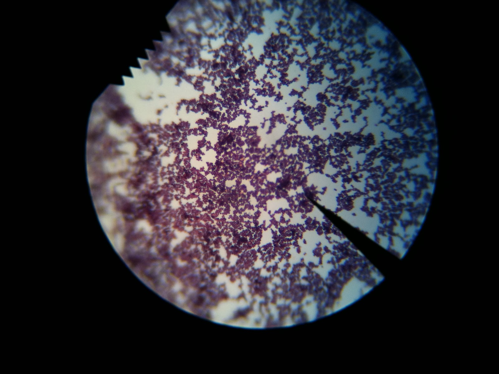
The next test performed on our bacterium told us whether each of them were gram-negative or gram-positive. Gram-positive bacteria have a much thicker layer of peptidoglycan in their cell wall, causing them to keep the first stain they receive much better than a gram-negative bacteria as shown below.

The next test performed on our bacterium told us whether each of them were gram-negative or gram-positive. Gram-positive bacteria have a much thicker layer of peptidoglycan in their cell wall, causing them to keep the first stain they receive much better than a gram-negative bacteria as shown below.

The first step of the staining process is to prepare a fixed smear of the bacteria as in past labs. Next, crystal violet is applied to the slide for 20 seconds.

After the smear is rinsed for excess dye, iodine is applied to the smear for one minute then rinsed again. The smear is then decolorized with 95% ethyl alcohol and rinsed again. As you can see in the chart above, the reactions of the two different types of bacteria at this point are already different. After decolorization, the color leaves a gram-negative bacteria but sticks in the gram-positive bacteria. This is why gram-positive bacteria have a purple/violet color to them and gram-negative bacteria do not. In the last step of the gram stain process, safranin is applied to the stain for a minute before one last rinse and a gentle blot to remove excess liquid.


The next lab, on September 22, 2011, we first looked closer at colony morphology and tried to identify our bacterium more accurately. We made simple stains of our bacteria and looked for shape, how/if they cluster, and the rest we had already accurately identified such as the raised edge.
Below is a similar colony morphology chart to the one in our books.

The result was that our unknown bacteria grows in spheres (it s a coccus bacteria - pictured above) and in tetrad clusters (pictured below), clusters of four, while our environmental bacteria grows in single spheres.

No comments:
Post a Comment